
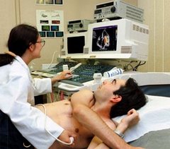
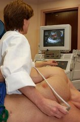
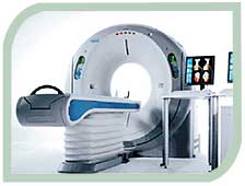
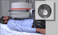
The advent of very advanced CAT scan imaging has enabled cardiologists to detect the earliest calcified and non-calcified plaque in coronary arteries. This noninvasive procedure has allowed the early detection of coronary artery disease before patients are stricken with sudden cardiac death and myocardial infarction (heart attack).
Westside Medical Imaging has been a pioneer and leader in the field of coronary imaging and 64 slice CT coronary angiography having performed nearly 4,000 scans to date and with that having an important impact on many lives. The use of 64 slice cardiac CT as a screening examination has been evaluated by Norman Lepor MD in a recent publication in the medical journal, Reviews in Cardiovascular Medicine.
Dr. Lepor is an Associate Clinical Professor of Medicine at the Geffen School of Medicine at UCLA and the co-director of Cardiovascular Imaging at Westside Medical Imaging. In this pivotal publication, Dr. Lepor describes the ability of this important technology to detect heart disease in the presymptomatic phase. In addition, he discusses the shortcoming of the current method of risk assessment using risk factor analysis which often underestimates the risk certain populations. Dr. Lepor describes in detail the risk and benefits of this approach.
He recommends that any person with one or more of the following heart attack risk factors including diabetes, smoker (past or current), high blood pressure, family history of heart disease, high cholesterol and age (> 45 years for males and > 50 years for females) be considered for this important screening examination. With the chance of having a heart attack much greater then developing breast cancer and colon cancer, coronary artery screening should save many lives.
See more about this incredible breakthrough at http://www.westsidemedimaging.com/
From PR Newswire


1 comment:
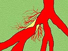
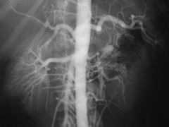
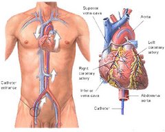
The process is minimally invasive, which means that there will be no major incision made on the body. The patient will be asked to lie down in the operation room. A mild sedative will be administered to him by intravenous means and the neck, arms and lower body will be made numb. Once the dye has entered the blood vessels, the x-ray machine will capture visual descriptions called fluoroscopy. The cardiologist (Heart doctor) may collect blood samples from your heart as well as assess the pressure of the blood flowing through the various parts of the body. The other important aspect is the oxygen content carried in the blood and supplied through out the human anatomy. Coronary heart diseases, congenital heart defects, probability of heart failure and other abnormalities can be comprehended through the process. Hospitals for Angiography in Thailand
Post a Comment